Abdominopelvic Regions and Quadrants Explained
Regions and Quadrants of the Abdominopelvic Cavity
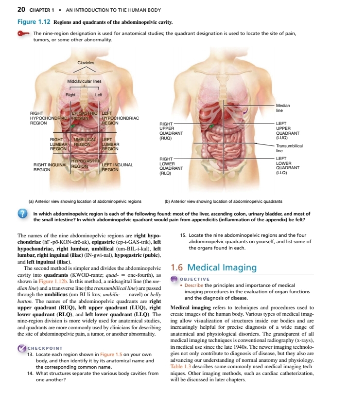
Overview
The image provides an anatomical reference of the abdominopelvic cavity, showing the nine abdominal regions and the four quadrants. These divisions are essential for identifying specific areas concerning medical issues, such as pain or abnormalities.
Abdominopelvic Regions
1. Nine Abdominopelvic Regions
-
Right Hypochondriac Region
- Location: Upper right section; contains parts of the liver and gallbladder.
-
Epigastric Region
- Location: Central upper section; primarily contains the stomach and parts of the liver.
-
Left Hypochondriac Region
- Location: Upper left section; includes parts of the spleen and stomach.
-
Right Lumbar Region
- Location: Middle right section; houses the ascending colon and parts of the small intestine.
-
Umbilical Region
- Location: Central region; includes the small intestine and part of the transverse colon.
-
Left Lumbar Region
- Location: Middle left section; contains the descending colon and kidneys.
-
Right Inguinal (Lateral) Region
- Location: Lower right section; encompasses the cecum and appendix.
-
Hypogastric (Pubic) Region
- Location: Central lower section; includes the urinary bladder and reproductive organs.
-
Left Inguinal (Lateral) Region
- Location: Lower left section; involves parts of the sigmoid colon.
2. Quadrants of the Abdominopelvic Cavity
- Right Upper Quadrant (RUQ)
- Contains: Most of the liver, gallbladder, and part of the colon.
- Left Upper Quadrant (LUQ)
- Contains: Most of the stomach, spleen, and part of the pancreas.
- Right Lower Quadrant (RLQ)
- Contains: Appendix, cecum, and part of the small intestine.
- Left Lower Quadrant (LLQ)
- Contains: Most of the small intestine and parts of the colon.
Medical Relevance
Understanding these regions and quadrants is critical for diagnosing medical conditions. For example:
- Appendicitis: Pain usually presents in the RLQ, indicating potential issues with the appendix.
- Liver Disease: Symptoms may manifest in the RUQ, where the liver is located.
Summary
The division of the abdominopelvic cavity into regions and quadrants aids in medical evaluation by providing a systematic approach to locate organs and identify the sources of pain or abnormalities. Familiarity with these anatomical divisions can enhance diagnostic accuracy and promote effective communication in clinical settings.
Reference:
Notes on Abdominopelvic Cavity and Membranes
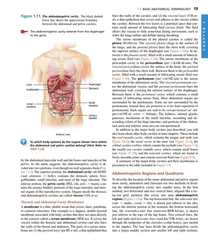
Abdominopelvic Cavity
-
Definition: The abdominopelvic cavity is the large body cavity that houses various organs.
- Thoughts: Understanding the layout of this cavity is essential in anatomy as it helps in localizing diseases and injuries.
- Additional Info: It is divided into two main sections: the abdominal cavity (upper portion) and the pelvic cavity (lower portion).
-
Boundary: The black dashed line in the image indicates the approximate boundary between the abdominal and pelvic cavities.
- Thoughts: Recognizing this boundary is crucial when discussing surgical procedures or conditions affecting either cavity.
- Additional Info: The diaphragm separates the thoracic cavity from the abdominopelvic cavity.
Major Organs in the Abdominopelvic Cavity
- Abdominal Cavity: Contains the liver, gallbladder, stomach, large intestine, small intestine, and urinary bladder.
- Thoughts: Each of these organs plays vital roles in digestion, metabolism, and waste elimination.
- Additional Info: The organization's study can help in understanding systemic diseases.
Serous Membranes
-
Pleura: The serous membrane associated with the lungs and diaphragm.
- Thoughts: This membrane reduces friction during breathing movements.
- Additional Info: It consists of two layers: parietal pleura (lines the cavity) and visceral pleura (covers the lungs).
-
Peritoneum: The serous membrane of the abdominal cavity.
- Thoughts: The peritoneum is significant for protecting abdominal organs and facilitating movement.
- Additional Info: It also has parietal (lining the cavity) and visceral (covering the organs) layers.
Thoracic and Abdominopelvic Cavities
- Membranes Role: Membranes like the peritoneum and pleura are crucial for enclosing organs and providing a lubricated surface for movement.
- Thoughts: Understanding the interaction between these membranes can illuminate several pathologies, including infections and adhesions.
- Additional Info: The presence of serous fluid in these cavities aids in organ functionality.
Abdominopelvic Regions and Quadrants
- Regions: The abdominopelvic cavity can be divided into quadrants for clinical purposes.
- Thoughts: Using quadrants helps in diagnosing abdominal issues by localizing pain or other symptoms.
- Additional Info: The first standard division is into four quadrants: right upper, left upper, right lower, and left lower quadrant.
| Quadrant | Location Description |
|---|---|
| Right Upper Quadrant | Contains liver, gallbladder |
| Left Upper Quadrant | Contains stomach, spleen |
| Right Lower Quadrant | Contains appendix, portions of the intestines |
| Left Lower Quadrant | Contains portions of the intestines, urinary bladder |
These notes aim to provide a comprehensive understanding of the anatomy of the abdominopelvic cavity and the importance of its components.
Notes on the Thoracic Cavity
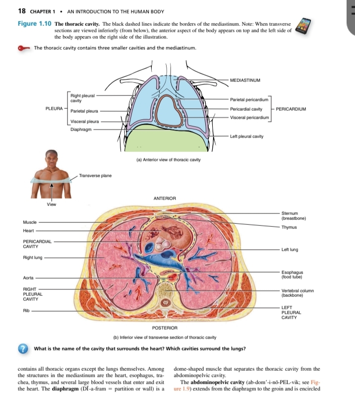
Overview of the Thoracic Cavity
- The thoracic cavity is a significant part of the human anatomy, housing vital organs such as the heart and lungs.
- It is divided into three smaller cavities: two pleural cavities and the mediastinum.
Components of the Thoracic Cavity
-
Pleural Cavities
- Right Pleural Cavity: Surrounds the right lung.
- Left Pleural Cavity: Surrounds the left lung.
- Visceral Pleura: The inner layer that directly covers the lungs.
- Parietal Pleura: The outer layer that lines the pleural cavity.
- Thoughts: The pleural cavities are crucial for respiratory mechanics, allowing the lungs to expand and contract.
-
Mediastinum
- Located between the pleural cavities, this area contains the heart, esophagus, trachea, thymus, and large blood vessels.
- Pericardial Cavity: A sub-cavity within the mediastinum specifically surrounding the heart. It consists of:
- Visceral Pericardium: The layer covering the heart.
- Parietal Pericardium: The outer layer that forms a sac around the heart.
- Thoughts: The mediastinum plays a vital role in separating and protecting the heart and great vessels from the lungs and chest wall.
-
Diaphragm
- A dome-shaped muscle forming the floor of the thoracic cavity, separating it from the abdominal cavity.
- Thoughts: The diaphragm is essential for respiration, as it contracts during inhalation, allowing air to be drawn into the lungs.
Summary of Cavity Functions
- Primary Functions: Protects the organs within, facilitates efficient breathing, and aids in blood circulation.
- The abdominopelvic cavity extends from the diaphragm down to the groin and is encapsulated by abdominal muscles.
Table: Thoracic Cavity Components
| Component | Description |
|---|---|
| Right Pleural Cavity | Surrounds the right lung |
| Left Pleural Cavity | Surrounds the left lung |
| Mediastinum | Contains the heart, esophagus, trachea, and thymus |
| Pericardial Cavity | Surrounds the heart |
| Visceral Pleura | Covers the lungs and heart |
| Parietal Pleura | Lines the pleural and pericardial cavities |
| Diaphragm | Muscle separating thoracic and abdominal cavities |
This organized summary outlines the essential components and functions of the thoracic cavity, highlighting its significance in human anatomy and physiology.
Reference:
Notes on Body Cavities
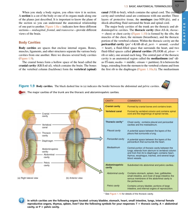
Anatomical Terminology
- Section View: When studying a body region, it is often analyzed in sections to understand relationships between different structures.
- Thoughts: Understanding sectional views is crucial for professions like medicine and anatomy studies as it provides clarity on organ placements and interactions.
Body Cavities
- Definition: Body cavities are spaces that contain and protect internal organs. They are separated by bones, muscles, ligaments, and other structures.
- Thoughts: Knowledge of body cavities is essential for comprehending how organs function in relation to one another and the overall body structure.
Major Body Cavities
- Cranial Cavity:
- Formed by cranial bones and contains the brain.
- Vertebral Canal:
- Formed by the vertebral column, contains the spinal cord and beginnings of spinal nerves.
Thoracic Cavity
- Components:
- Contains the pleural (lung), pericardial (heart), and mediastinum regions.
- Thoughts: Understanding the thoracic cavity is key in fields like cardiology and pulmonology, as they focus on heart and lung health.
Abdominopelvic Cavity
- Subdivision:
- Divided into abdominal and pelvic cavities.
| CAVITY | COMMENTS |
|---|---|
| Cranial cavity | Formed by cranial bones and contains the brain. |
| Vertebral canal | Formed by the vertebral column and contains spinal cord and beginnings of spinal nerves. |
| Thoracic cavity | Contains pleural and pericardial cavities and the mediastinum. |
| Pleural cavity | Potential space between the layers of the pleura that surrounds a lung. |
| Pericardial cavity | Potential space between the layers of the pericardium that surrounds the heart. |
| Mediastinum | Central portion of thoracic cavity between the lungs, extending from the sternum to the vertebral column. |
| Abdominal cavity | Contains stomach, liver, gallbladder, small intestine, and part of large intestine. |
| Pelvic cavity | Contains urinary bladder, portions of the large intestine, and internal organs of reproduction. |
Quiz Question
- In which cavities are the following organs located: urinary bladder, stomach, heart, small intestine, lungs, internal female reproductive organs, thymus, spleen, liver?
- Answer Format: Use the symbols: T = thoracic cavity, A = abdominal cavity, P = pelvic cavity.
Reference:
Notes on Human Body Planes and Sections
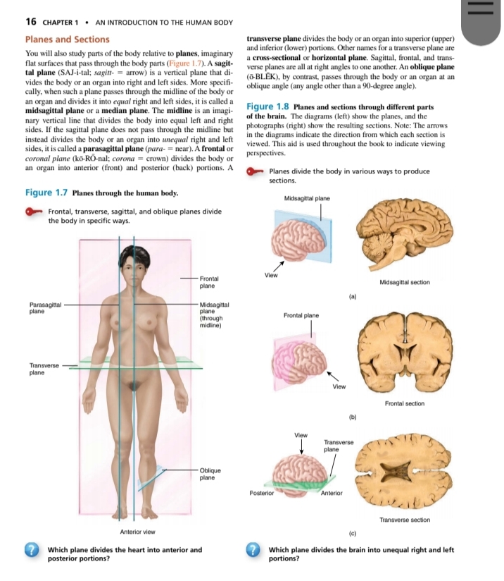
Planes and Sections
-
Sagittal Plane:
- Imaginary vertical plane that divides the body into right and left sides.
- More specifically referred to as a midsagittal plane if it divides the body into equal halves. When it divides into unequal parts, it is called a parasagittal plane.
- Understanding the sagittal plane is crucial for anatomical studies, especially in observing bilateral symmetry in organisms.
-
Frontal Plane:
- Also known as the coronal plane; it bisects the body into anterior (front) and posterior (back) portions.
- This plane is essential for understanding different anatomical views, especially in imaging techniques like MRIs, where front-to-back perspectives are essential.
-
Transverse Plane:
- This plane divides the body or an organ into superior (upper) and inferior (lower) portions.
- It is also referred to as a horizontal or cross-sectional plane.
- A good grasp of the transverse plane is essential for surgical planning and procedures that involve horizontal sections of anatomy.
-
Oblique Plane:
- This plane cuts through the body at an angle, providing a different perspective compared to the standard planes.
- Understanding oblique planes can help visualize complex structures in 3D, which is often necessary in advanced imaging techniques.
Brain Sections
- Different sections of the brain can be visualized using the planes:
- Midsagittal Section: Shows symmetrical left and right halves of the brain.
- Frontal Section: Demonstrates what lies in the anterior and posterior aspects of the brain.
- Transverse Section: Reveals additional internal structures by slicing horizontally.
Quick Reference Table: Planes of the Body
| Plane Type | Description | Sections Resulting |
|---|---|---|
| Sagittal Plane | Divides body into right and left parts | Midsagittal, Parasagittal |
| Frontal Plane | Divides body into anterior and posterior parts | - |
| Transverse Plane | Divides body into superior and inferior parts | - |
| Oblique Plane | Divides body at an angle | - |
Questions
-
Which plane divides the heart into anterior and posterior portions?
- The frontal (coronal) plane.
-
Which plane divides the brain into unequal right and left portions?
- The parasagittal plane.
Reference:
Directional Terms in Anatomy
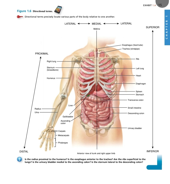
This image illustrates various directional terms used to locate parts of the human body relative to one another. Understanding these terms is crucial for accurately describing the locations of structures and functions within the body.
Key Directional Terms:
-
Lateral: Refers to the sides of the body or structures away from the midline.
- Thoughts: The use of lateral terms helps in identifying injuries or conditions affecting specific areas, such as lateral epicondylitis (tennis elbow).
-
Medial: Indicates structures closer to the midline of the body.
- Additional Info: Knowing the medial positions can aid in diagnosing conditions that may affect parts of the body that are closer to the center.
-
Proximal: Describes a position closer to the point of attachment or origin.
- Thoughts: This term is often used in describing limbs, as it helps establish the closeness of different segments, such as the proximal radius relative to the wrist.
-
Distal: Opposite of proximal; refers to a position further from the point of attachment.
- Additional Info: Understanding this can help in surgery or medical treatments, as physicians need to know the extent of tissue removal or damage.
-
Superior: Denotes a position above or higher than another structure.
- Thoughts: Knowing which organs are superior helps in surgical planning and emergency medical situations.
-
Inferior: Indicates a position lower than another structure.
- Additional Info: This clarity is useful in various medical scenarios, such as when discussing abdominal organs.
Relevant Structures in the Image:
| Structure | Position Relation |
|---|---|
| Right Lung | Lateral |
| Sternum (breastbone) | Medial |
| Humerus | Proximal to Radius |
| Liver | Inferior to Diaphragm |
| Gallbladder | Medial to Ascending Colon |
| Urinary Bladder | Inferior to Descending Colon |
Thought-Provoking Questions:
-
Is the radius proximal to the humerus?
- Answer: Yes, because the radius is closer to the elbow joint compared to the wrist.
-
Is the esophagus anterior to the trachea?
- Answer: No, the trachea is located anteriorly to the esophagus.
-
Are the ribs superficial to the lungs?
- Answer: Yes, ribs provide a protective barrier and are external to the lungs.
-
Is the urinary bladder medial to the ascending colon?
- Answer: Yes, the urinary bladder is positioned closer to the midline than the ascending colon.
-
Is the sternum lateral to the descending colon?
- Answer: Yes, the sternum (breastbone) is located towards the front (anterior) and to the side relative to the descending colon.
These notes provide an organized approach to the directional terms and structures related to human anatomy. Understanding these concepts is vital for students and professionals in medical and health-related fields.
Reference:
Directional Terms in Anatomy
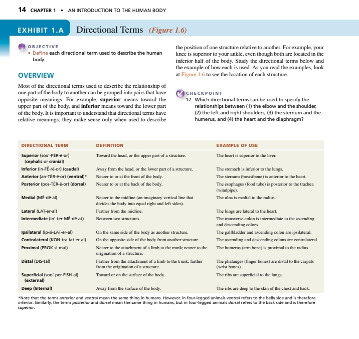
Overview of Directional Terms
- Directional terms describe the relationship between different parts of the body.
- They often come in pairs with opposite meanings (e.g., superior vs. inferior).
Directional Terms Table
| Directional Term | Definition | Example of Use |
|---|---|---|
| Superior | Toward the head, or the upper part of a structure | The heart is superior to the lungs. |
| Inferior | Away from the head, or the lower part of a structure | The stomach is inferior to the lungs. |
| Anterior | Nearer to the front of the body | The sternum (breastbone) is anterior to the heart. |
| Posterior | Nearer to or at the back of the body | The esophagus (food tube) is posterior to the trachea. |
| Medial | Nearer to the midline of the body | The knee is medial to the ankle. |
| Lateral | Farther from the midline | The lungs are lateral to the heart. |
| Intermediate | Between two structures | The transverse colon is intermediate to the ascending and descending colons. |
| Ipsilateral | On the same side of the body as another structure | The gallbladder and ascending colon are ipsilateral. |
| Contralateral | On the opposite side of the body from another structure | The ascending and descending colons are contralateral. |
| Proximal | Nearer to the attachment of a limb | The humerus (arm bone) is proximal to the radius. |
| Distal | Farther from the origin of a structure | The phalanges (finger bones) are distal to the carpals. |
| Superficial | Toward or on the surface of the body | The ribs are superficial to the skin of the chest. |
| Deep | Away from the surface of the body | The ribs are deep to the skin of the chest and back. |
Key Concepts
- Superior vs. Inferior: Understanding these terms helps in identifying the positioning of organs in relation to one another.
- Anterior vs. Posterior: Important for describing the front and back orientation of the body.
- Medial vs. Lateral: Useful for assessing proximity to the midline and understanding bilateral symmetry.
Additional Information
- Directional terms are crucial in fields like medicine, anatomy, and physical therapy. They provide clarity and precision when discussing anatomy, especially during surgical procedures or diagnostic assessments.
- These terms also highlight the importance of relative position, which is essential for understanding biological function and structure.
Reference:
Anatomical Position and Terminology
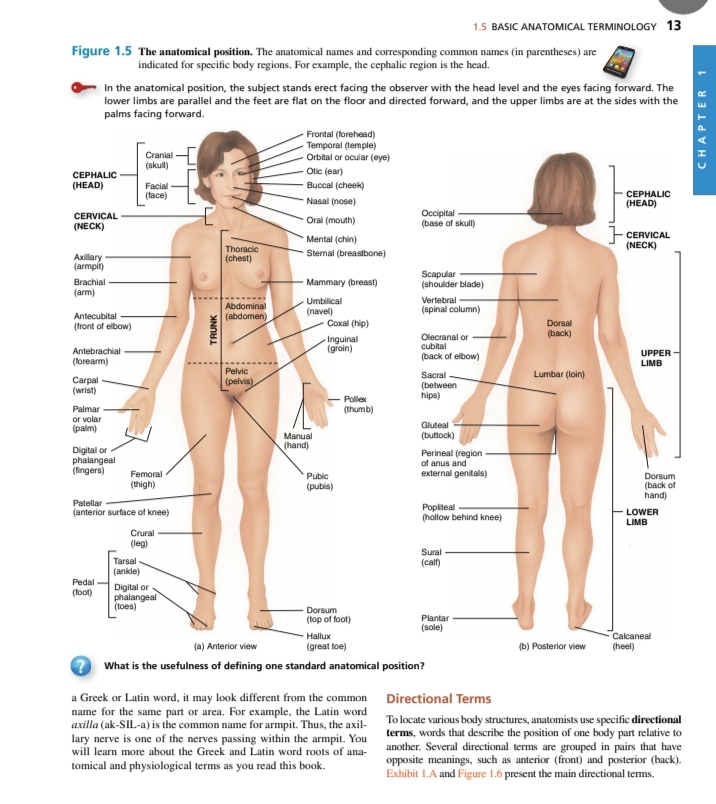
The Anatomical Position
- Definition: The anatomical position is a standardized way of observing or describing the human body. In this position, the person stands upright, facing forward, with arms at the sides and palms facing forward. This position serves as the reference point for anatomical terminology.
- Importance: Understanding the anatomical position is crucial as it provides a clear framework for identifying locations and relationships between different body parts.
Body Regions and Corresponding Terms
Regions Listed in the Image
| Body Region | Anatomical Term | Common Name |
|---|---|---|
| Head | Cephalic | Skull |
| Neck | Cervical | Neck |
| Chest | Thoracic | Chest |
| Abdomen | Abdominal | Abdomen |
| Arm | Brachial | Arm |
| Armpit | Axillary | Armpit |
| Forearm | Antebrachial | Front of forearm |
| Wrist | Carpal | Wrist |
| Hand | Manual | Hand |
| Leg | Crural | Leg |
| Thigh | Femoral | Thigh |
| Knee | Patellar | Anterior surface of knee |
| Ankle | Tarsal | Ankle |
| Foot | Pedal | Foot |
| Bottom of Foot | Plantar | Sole |
Thoughts on Body Regions:
- Utility of Terminology: Using specific anatomical terms facilitates clear communication in medical, educational, and research settings. For instance, knowing the difference between "brachial" (arm) and "crural" (leg) can prevent misunderstandings during discussions related to anatomy or health.
- Common Names vs. Anatomical Terms: While common names may seem simpler, anatomical terms are precise and often derived from Latin or Greek, providing a universal language for anatomists and healthcare professionals.
Directional Terms
- Purpose: Directional terms are used to describe the position of one body part in relation to another, enhancing clarity in anatomical descriptions.
- Examples: Terms such as anterior (front) and posterior (back), superior (above) and inferior (below) help create a comprehensive understanding of human anatomy and its organization.
Summary of Key Directional Terms (not in table):
- Anterior (Ventral): Front
- Posterior (Dorsal): Back
- Superior: Above
- Inferior: Below
- Medial: Toward the midline
- Lateral: Away from the midline
- Proximal: Closer to the point of attachment
- Distal: Further from the point of attachment
Conclusion
Understanding the anatomical position and related terminology is fundamental for anyone studying or working in health-related fields. This knowledge aids in precise communication and serves as a foundation for more advanced studies in anatomy and physiology.
Reference:
Notes on Introduction to the Human Body
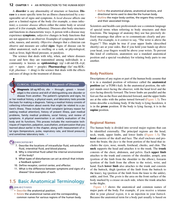
Definition of Disease
- Disorder: An abnormality of structure or function.
- Disease: A specific term for an illness characterized by recognizable signs and symptoms.
- Local disease: Affects a particular region of the body (e.g., influenza).
- Systemic disease: Affects the entire body or several parts.
Thoughts:
Understanding the distinction between different types of diseases helps in diagnosis and treatment planning.
Epidemiology and Pharmacology
- Epidemiology: The study of how and when diseases occur and are transmitted among individuals.
- Pharmacology: The science that deals with the effects and uses of drugs in the treatment of disease.
Thoughts:
Knowledge of these fields is crucial for health professionals to understand disease patterns and therapeutic interventions.
Anatomical Terms
- Anatomical position: The standard reference position for the body.
- Observer stands facing the subject, with the upper limbs at the sides and palms facing forward.
- If the body is facing upwards, it is in the supine position.
Thoughts:
This standard position is essential for clear communication in the medical field regarding body positioning.
Body Positions
- Descriptions of body regions assume the anatomical position for consistency.
- Variations in positions can lead to confusion in anatomical references.
Thoughts:
Learning these terms can assist in understanding medical literature and clinical communication.
Regional Names
- Major regions: Head, trunk, upper limbs, and lower limbs.
- Face: The front part of the head including the eyes, nose, mouth, and cheeks.
- Upper Limbs: Consist of shoulder, arm, forearm, wrist, and hand.
- Lower Limbs: Include the thigh, leg, ankle, and foot.
Thoughts:
Familiarity with regional names helps students and professionals locate and describe specific parts of the human body efficiently.
Checkpoints
- Fluid compartments: Understand the locations of intracellular fluid, extracellular fluid, interstitial fluid, and blood plasma.
- Interstitial fluid role: Recognize how it facilitates the internal environment.
- Disturbances and feedback: Learn how disturbances can stimulate feedback systems.
Thoughts:
These checkpoints reinforce knowledge necessary for both clinical practice and understanding human physiology.
Reference:
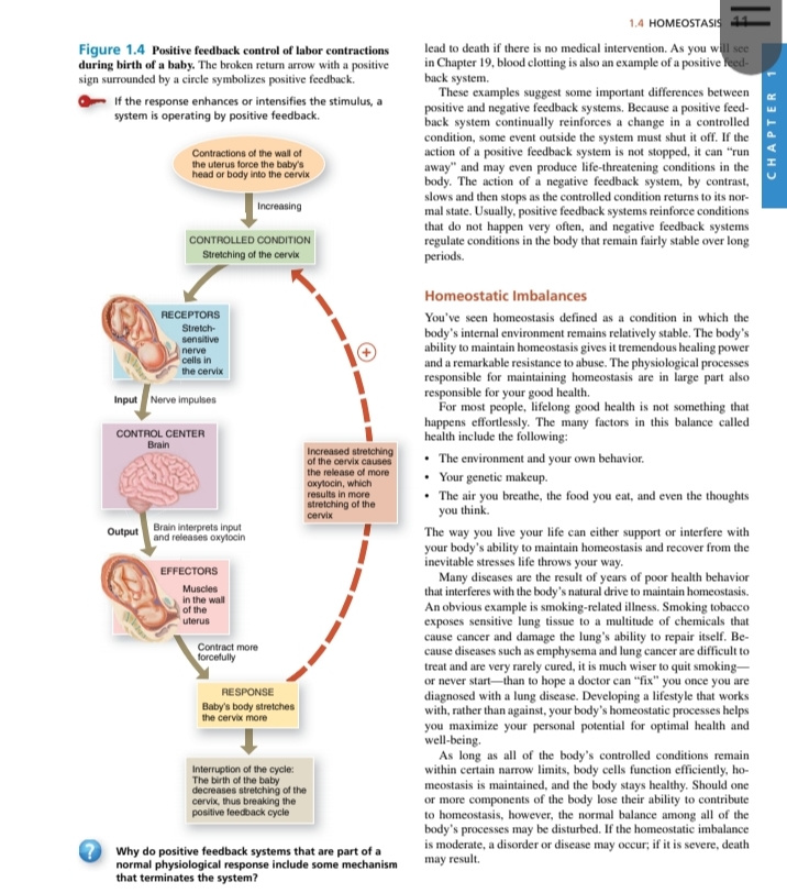
Positive Feedback Control of Labor Contractions
Key Points
-
Positive Feedback Mechanism
- The example of labor contractions in childbirth demonstrates a positive feedback loop where an initial stimulus (the baby's head pressing against the cervix) leads to a response (increased contractions).
- Thoughts: This mechanism shows how systems can amplify a process, which is crucial in situations like childbirth where rapid response and escalation are necessary.
-
Components of the Feedback Loop
-
Receptors: Stretch-sensitive cells in the cervix detect the stretching.
- Additional Info: These receptors are critical for initiating the feedback loop by sending nerve impulses to the control center when the cervix is stretched.
-
Control Center: The brain interprets inputs and releases oxytocin, a hormone that causes contractions.
- Thoughts: Oxytocin plays a vital role in childbirth, promoting strong contractions that aid in delivery.
-
Effectors: Muscles in the uterine wall contract in response to increased oxytocin levels.
- Additional Info: These contractions help to push the baby further down the birth canal, causing more stretching of the cervix, which in turn triggers more oxytocin release.
-
Response: The baby's position continues to stretch the cervix, perpetuating the cycle until delivery occurs.
- Thoughts: This cycle is crucial for ensuring the timely and effective process of labor.
-
-
Interruption of the Cycle
- The cycle continues until interrupted by the birth of the baby, effectively terminating the feedback loop.
- Additional Info: Once the baby is born, the stimulus that initiated the feedback response is removed, stopping the release of oxytocin.
- The cycle continues until interrupted by the birth of the baby, effectively terminating the feedback loop.
Homeostatic Imbalances
-
Definition of Homeostasis
- Homeostasis refers to the body's ability to maintain a stable internal environment despite external changes.
- Thoughts: Understanding this concept is fundamental to recognizing how various physiological processes interact to preserve health.
- Homeostasis refers to the body's ability to maintain a stable internal environment despite external changes.
-
Importance of Homeostasis
- The body's internal environment remains stable through numerous physiological responses that are crucial for health.
- Additional Info: Disruptions in homeostasis can lead to significant health issues, highlighting the importance of adaptive and regulatory mechanisms.
- The body's internal environment remains stable through numerous physiological responses that are crucial for health.
-
Factors Influencing Homeostasis
- Environment and Behavior: Lifestyle choices such as diet and exercise can greatly impact the body's balance.
- Genetic Makeup: Genetic predispositions play a role in how an individual's body manages stress and disease.
- External Factors: Elements like air quality and nutrition directly influence homeostasis.
-
Consequences of Disruption
- Imbalances can lead to disorders where the body struggles to heal or maintain normal function, leading to severe health issues.
- Thoughts: Recognizing early signs of imbalance can be vital for preventative healthcare.
- Imbalances can lead to disorders where the body struggles to heal or maintain normal function, leading to severe health issues.
| Component | Description |
|---|---|
| Receptors | Stretch-sensitive cells in the cervix. |
| Control Center | Brain interprets the signal and releases oxytocin. |
| Effectors | Muscles in the uterine wall contract in response to oxytocin. |
| Response | The baby's position stretches the cervix, perpetuating the feedback cycle. |
| Cycle Interruption | The cycle is interrupted by the birth of the baby, stopping the feedback loop. |
Reference:
Homeostasis and Feedback Systems
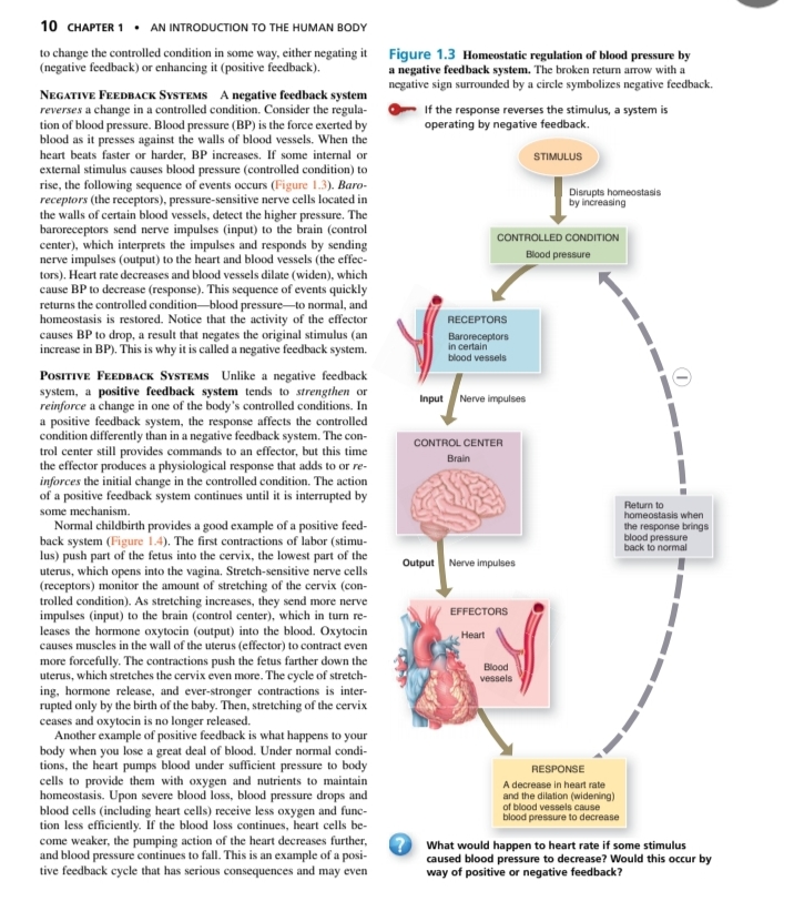
Negative Feedback Systems
- Definition: A negative feedback system reverses a change in a controlled condition.
- Example: Blood Pressure Regulation
- Process:
- Stimulus: High blood pressure detected by baroreceptors.
- Receptors: Baroreceptors send nerve impulses to the brain (control center).
- Control Center: Brain interprets signals and responds by sending nerve impulses to effectors.
- Effectors: Heart decreases rate, blood vessels dilate (widen).
- Response: Blood pressure decreases back to normal.
- Process:
- Example: Blood Pressure Regulation
- Importance: This system maintains homeostasis by keeping physiological parameters within a narrow range.
Positive Feedback Systems
- Definition: A positive feedback system amplifies a change in a controlled condition.
- Example: Normal Childbirth
- Process:
- Stimulus: Contractions push the fetus into the cervix.
- Receptors: Stretch-sensitive nerve cells monitor the stretching of the cervix.
- Control Center: Brain receives nerve impulses.
- Effectors: Brain releases oxytocin into the blood.
- Response: Oxytocin intensifies contractions, pushing the fetus further down.
- Process:
- Example: Normal Childbirth
- Importance: Positive feedback is usually temporary and must be interrupted for homeostasis to be restored.
Comparison of Feedback Systems
| Feature | Negative Feedback | Positive Feedback |
|---|---|---|
| Direction of Response | Reverses change | Reinforces change |
| Control Mechanism | Maintains stability | Drives processes in one direction |
| Typical Outcome | Homeostasis | Completion of a process (e.g., childbirth) |
Thought Process
- Negative Feedback Systems: Essential for regular physiological functioning. For instance, if blood pressure remains consistently high without regulation, it may lead to health issues such as hypertension.
- Positive Feedback Systems: Typically seen in processes that need to be pushed to completion, such as childbirth or blood clotting. They must be finely tuned to avoid excessive reactions that could lead to harm, like prolonged contractions that could endanger both mother and baby.
Reference:
Control of Homeostasis
Introduction to Homeostasis
- Definition: Homeostasis is the process by which the human body maintains a stable internal environment despite external changes.
- Importance: Essential for survival; disruption can lead to health problems.
Sources of Disruption
- External Factors: Includes environmental stressors such as extreme temperatures or lack of nutrition (e.g., low oxygen or blood glucose levels).
- Internal Factors: Psychological stress or physiological conditions can also disrupt homeostasis.
Regulatory Systems
- Nervous System: Responds quickly to maintain homeostasis through nerve impulses that act on various organs.
- Endocrine System: Works more slowly by sending hormones into the bloodstream, affecting long-term changes (e.g., growth, metabolism).
Key Concepts
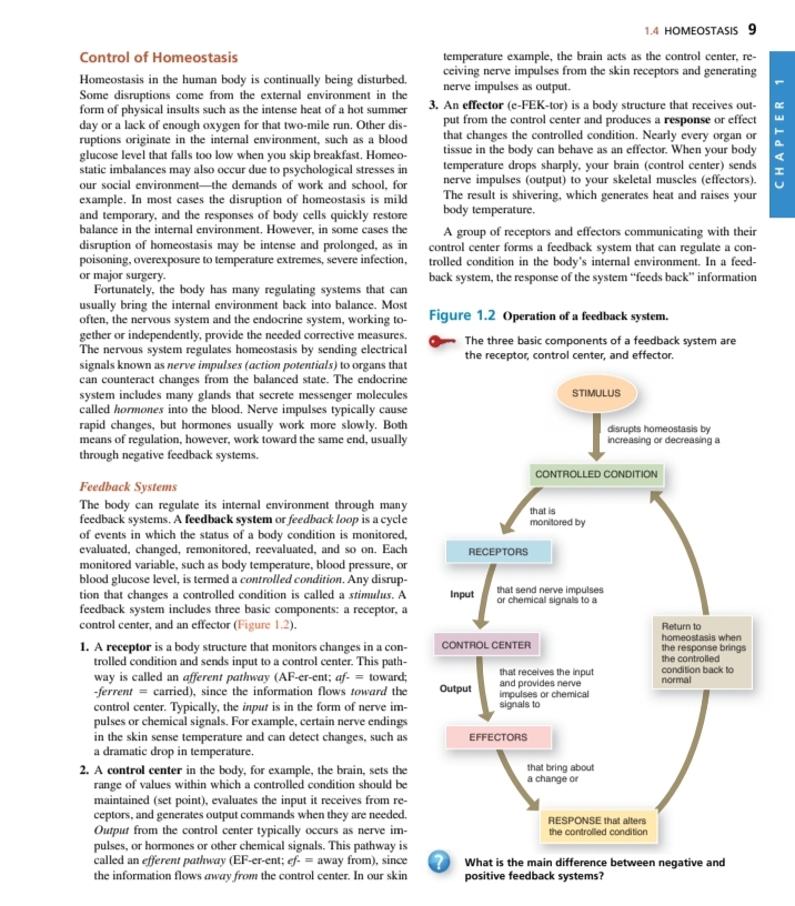
- Feedback Systems: The body utilizes feedback loops to monitor and adjust its internal environment.
- Types: Primarily negative feedback, which counteracts changes, and positive feedback, which amplifies changes.
Components of a Feedback System
-
Receptors: Monitor changes in a controlled condition and send information to the control center.
- Example: Baroreceptors in blood vessels detect changes in blood pressure.
-
Control Center: Receives input from receptors and determines the appropriate response.
- Example: The brain processes information from receptors and decides how to act.
-
Effectors: Execute the response to bring about a change in the controlled condition.
- Example: Muscles or organs respond by changing their activity (e.g., heart rate adjustments).
Feedback Loop Process
- Stimulus: Any factor that disrupts homeostasis (e.g., changes in temperature).
- Input: Signals sent from receptors to the control center.
- Output: Commands sent from the control center to effectors.
- Response: Changes enacted by effectors to restore homeostasis.
Summary of Feedback Systems
- Negative Feedback: Reduces the output or activity to return to stability (e.g., temperature control).
- Positive Feedback: Increases output or activity to enhance the process (e.g., blood clotting).
Diagram Reference
- Figure 1.2 illustrates the operation of a feedback system, showing the interactions between stimulus, receptors, control center, and effectors, culminating in a response that maintains stability.
Conclusion
Understanding the control of homeostasis is vital for recognizing how the body responds to internal and external changes, emphasizing the delicate balance that sustains life.
Reference: