Understanding Passive Transport: Diffusion Mechanisms
Plasma Membrane and Transport Mechanisms
Fluid Mosaic Model
-
Composition: The plasma membrane consists of lipids and proteins. The lipids form a fluid structure with proteins embedded, creating a mosaic pattern.
- Insight: This model highlights the dynamic nature of the membrane, allowing for flexibility and movement.
- Additional Info: The term "fluid mosaic model" was proposed by Singer and Nicolson in 1972.
-
Lipid Arrangement: Lipids are organized in two layers: an outer layer and an inner layer. Each lipid molecule has a hydrophilic head and a hydrophobic tail.
- Insight: This arrangement is crucial for forming a barrier that separates the cell from its environment.
- Additional Info: The hydrophilic heads face outward, interacting with water, while the hydrophobic tails face inward, avoiding water.
-
Protein Types: Proteins in the membrane are globular and categorized into:
- Peripheral (Extrinsic) Proteins: Loosely bound to the surface.
- Integral (Intrinsic) Proteins: Embedded within the lipid bilayer.
- Insight: Integral proteins play a key role in transport and communication across the membrane.
- Additional Info: Glycoproteins and glycolipids are formed when proteins and lipids attach to sugars.
-
Membrane Dynamics: Lipid molecules are not fixed and can move laterally, allowing proteins to move as well.
- Insight: This fluidity is essential for membrane function, including cell signaling and transport.
-
Glycocalyx: A sugar coating formed by carbohydrates in intrinsic proteins and glycolipids, recognizing foreign proteins and protecting the cell.
- Insight: The glycocalyx is important for immune response and cell recognition.
Transport Across Membranes
-
Function: The plasma membrane regulates material flow in and out of the cell, a process known as transport.
- Insight: This regulation is vital for maintaining homeostasis within the cell.
-
Selective Permeability: The membrane is semi-permeable, allowing certain substances to pass while blocking others.
- Insight: This property is crucial for nutrient uptake and waste removal.
Types of Transport
-
Passive Transport: Does not require energy.
- Simple Diffusion: Movement from high to low concentration until equilibrium is reached.
- Insight: Simple diffusion is driven by the concentration gradient and does not require cellular energy (ATP).
- Simple Diffusion: Movement from high to low concentration until equilibrium is reached.
-
Active Transport: Requires energy to move substances against their concentration gradient.
- Primary Active Transport: Direct use of ATP to transport molecules.
- Secondary Active Transport: Uses the energy from the electrochemical gradient created by primary active transport.
-
Transport Mechanisms:
- Passive Transport:
- Simple Diffusion
- Facilitated Diffusion
- Active Transport:
- Primary Active Transport
- Secondary Active Transport
- Passive Transport:
-
Additional Info: Facilitated diffusion involves transport proteins to help move substances across the membrane without energy.
These notes provide a comprehensive overview of the plasma membrane's structure and function, emphasizing the fluid mosaic model and various transport mechanisms.
Passive Transport and Diffusion
Overview
The movement of molecules across the plasma membrane from a region of higher concentration to a region of lower concentration is called passive transport. This process does not require energy input from the cell.
i) Simple Diffusion
-
Definition: Simple diffusion is the passive movement of solute from a high concentration to a lower concentration until equilibrium is reached.
- Insight: This process is driven by the concentration gradient and does not require cellular energy (ATP).
- Additional Information: Simple diffusion occurs directly through the lipid bilayer without the need for membrane proteins.
-
Characteristics:
- Does not require membrane proteins.
- Movement is from higher to lower concentration.
- Achieves equilibrium when concentrations are uniform.
-
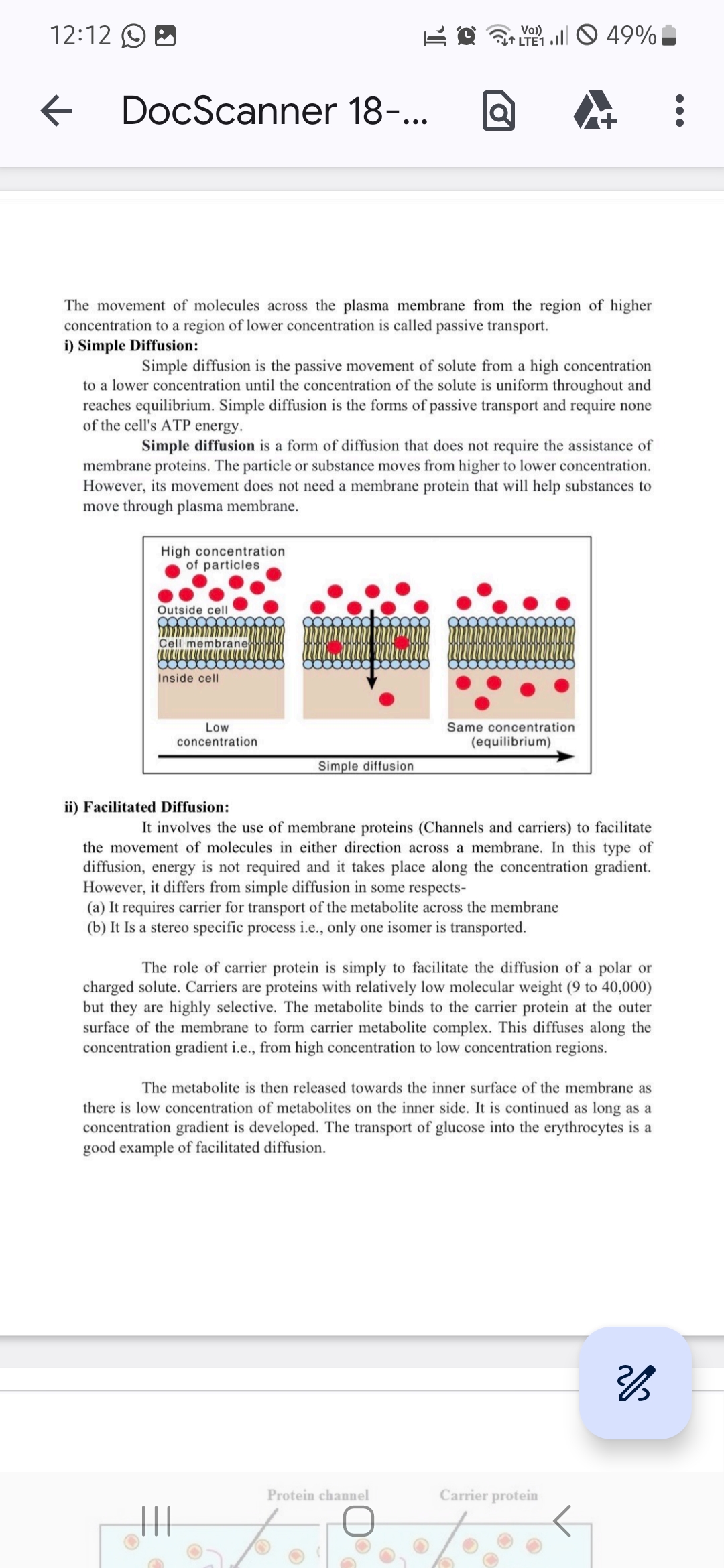
Diagram Explanation:
- The diagram illustrates particles moving from an area of high concentration outside the cell to a lower concentration inside the cell until equilibrium is reached.
ii) Facilitated Diffusion
-
Definition: Facilitated diffusion involves the use of membrane proteins (channels and carriers) to assist the movement of molecules across the membrane.
- Insight: This process is also passive and does not require energy, but it is necessary for molecules that cannot diffuse directly through the lipid bilayer.
- Additional Information: Facilitated diffusion is specific to certain molecules and involves protein channels or carriers.
-
Characteristics:
- Requires carrier proteins for transport.
- Is a stereo-specific process, meaning only specific isomers are transported.
- Proteins involved have a low molecular weight (9 to 40,000) and are highly selective.
-
Process:
- The metabolite binds to the carrier protein on the membrane's outer surface.
- A metabolite complex is formed and diffuses along the concentration gradient.
- The metabolite is released on the inner surface where the concentration is lower.
-
Example:
- The transport of glucose into erythrocytes (red blood cells) is a classic example of facilitated diffusion.
-
Diagram Explanation:
- The diagram shows how protein channels and carrier proteins facilitate the movement of particles across the membrane.
These notes provide a comprehensive understanding of passive transport mechanisms, focusing on simple and facilitated diffusion, their characteristics, and their biological significance.
Cell Junctions and Their Functions
1. Gap Junctions
Overview
- Also Known As: Communicating junctions, macula communicans, nexuses.
- Function: Allow direct passage of molecules between two cells.
Structure
- Composition: Transmembrane channels called pores, arranged closely.
- Variability: Number of gap junctions between cells can vary.
Location
- Found In: Epithelia, nerves, cardiac muscle, smooth muscle (e.g., intestines).
- Role: Coordinate activity of adjacent cells, e.g., electrical signal transmission in heart cells.
Detailed Structure
- Channel Formation: Made of two half channels (hemichannels) from each cell's membrane.
- Connexon: Each half channel is a connexon, composed of six connexins (protein units).
- Protein Arrangement: 12 circularly arranged protein units per channel.
2. Desmosomes
Overview
- Also Known As: Maculae adherentes.
- Function: Act as rivets through the plasma membrane of adjacent cells.
Structure
- Intermediate Filaments: Composed of keratin or desmin, attached to membrane-associated proteins.
- Cadherin Molecules: Form anchors by attaching to cytoplasmic plaques and binding to cadherins of adjacent cells.
Hemidesmosomes
- Function: Link cytoskeleton to extracellular matrix components like basal laminae.
- Difference from Desmosomes: Use integrins instead of cadherins for transmembrane anchors.
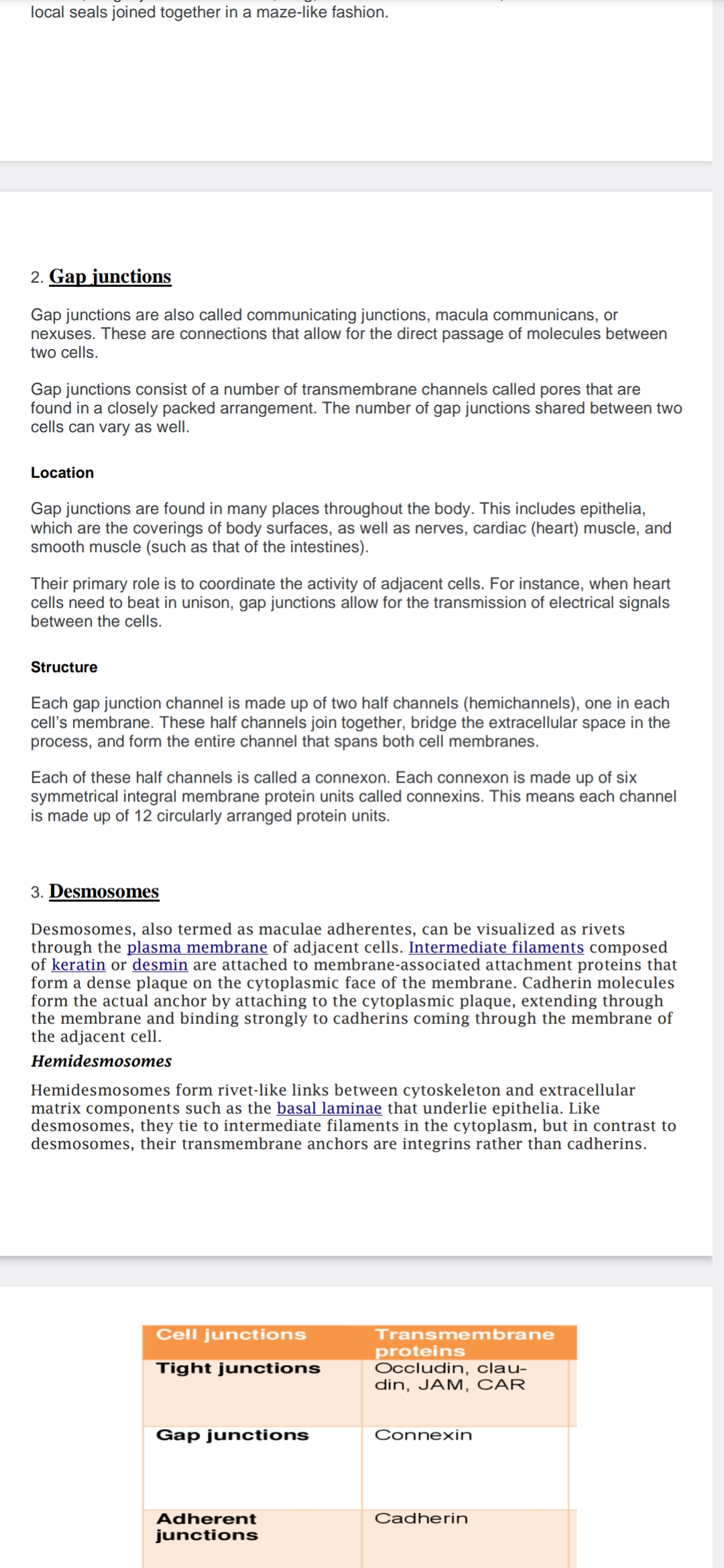
3. Cell Junctions and Transmembrane Proteins
| Cell Junctions | Transmembrane Proteins |
|---|---|
| Tight junctions | Occludin, claudin, JAM, CAR |
| Gap junctions | Connexin |
| Adherent junctions | Cadherin |
Insights
- Tight Junctions: Create a seal to prevent passage of molecules between cells.
- Gap Junctions: Facilitate communication and coordination between cells.
- Adherent Junctions: Provide mechanical stability by linking cytoskeletons of adjacent cells.
Additional Information
- Connexins: Essential for forming gap junctions, allowing ion and molecule passage.
- Cadherins: Calcium-dependent adhesion proteins crucial for cell-cell adhesion.
- Integrins: Transmembrane receptors that facilitate cell-ECM adhesion in hemidesmosomes.
These notes provide a comprehensive understanding of cell junctions, their structures, functions, and associated proteins, highlighting their roles in cellular communication and adhesion.
Cell Junctions: Tight Junctions, Gap Junctions, Desmosomes
Overview
Cell Junctions (Intercellular Bridges):
- Structures that provide contact or adhesion between neighboring cells or between a cell and the extracellular matrix in animals.
- Composed of multiprotein complexes.
- Essential for maintaining the paracellular barrier of epithelia and controlling paracellular transport.
- Abundant in epithelial tissues, aiding in holding animal cells together.
- Facilitate communication between cells via specialized protein complexes, reducing stress on cells.
Types of Cell Junctions:
- Tight Junctions
- Gap Junctions
- Desmosomes
In Plants and Fungi:
- Similar communication channels are known as plasmodesmata in plants and septal pores in fungi.
Tight Junctions
Composition
- Symmetrical Cell Junctions: Composed of proteins that connect the junction to and between cells.
- Key Proteins:
- Occludins: Maintain the barrier between adjacent cells.
- Claudins: Form the backbone of tight junction strands.
- Junctional Adhesion Molecules (JAMs): Immunoglobulin proteins that seal intercellular space.
- Zonula Occludens (ZO): Link tight junctions to the cell's cytoskeleton.
Structure
- Not a continuous seal; resembles a series of local seals joined in a maze-like pattern.
Insights
- Occludins and Claudins: Major components that ensure the integrity of tight junctions.
- Function: Prevents the passage of molecules and ions through the space between cells, maintaining distinct environments on either side of the epithelium.
Additional Information
- Paracellular Transport: Movement of substances across an epithelium by passing through the intercellular space between cells.
- Cell Adhesion Molecules: Proteins located on the cell surface involved in binding with other cells or with the extracellular matrix.
- Extracellular Matrix: A network of non-living tissue that provides support to cells.
Understanding cell junctions is crucial for comprehending how cells interact, communicate, and maintain structural integrity within tissues.